Акс:Trophozoites of Entamoeba histolytica with ingested erythrocytes.JPG
Нусхаи ҳаҷман ва сифатан баландтар дастрас нест.
Trophozoites_of_Entamoeba_histolytica_with_ingested_erythrocytes.JPG ((282 × 198 пиксел, ҳаҷми парванда: 27 кбайт, навъи MIME: image/jpeg))
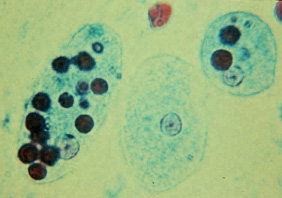
| Тавсифот | Trophozoites of Entamoeba histolytica with ingested erythrocytes (trichrome stain). The ingested erythrocytes appear as dark inclusions. Erythrophagocytosis is the only characteristic that can be used to differentiate morphologically E. histolytica from the nonpathogenic E. dispar. In these specimens, the parasite nuclei have the typical small, centrally located karyosome, and thin, uniform peripheral chromatin. | |||
| Манбаъ | DPD CDC http://www.dpd.cdc.gov/dpdx/images/ParasiteImages/A-F/Amebiasis/E_histol_trophs_F.JPG | |||
| Муаллиф | ||||
| Иҷозат (Пешроҳандозии ин парванда) |
|
Таърихи файл
Рӯи таърихҳо клик кунед то нусхаи марбути парвандаро бубинед.
| Таърих | Бандангуштӣ | Андоза | Корбар | Тавзеҳ | |
|---|---|---|---|---|---|
| нусхаи феълӣ | 13:38, 29 апрели 2006 | | 282 × 198 (27 кбайт) | Patho | {{Information| |Description= Trophozoites of Entamoeba histolytica with ingested erythrocytes (trichrome stain). The ingested erythrocytes appear as dark inclusions. Erythrophagocytosis is the only characteristic that can be used to differentiate morpho |
Пайвандҳо
Саҳифаҳои зерин ба ин акс пайванданд:
Истифодаи саросарии парванда
Викиҳои дигари зерин ин файлро истифода мекунанд:
- Истифода дар ar.wikipedia.org
- Истифода дар bn.wikipedia.org
- Истифода дар bs.wikipedia.org
- Истифода дар ca.wikipedia.org
- Истифода дар cs.wikipedia.org
- Истифода дар da.wikipedia.org
- Истифода дар de.wikipedia.org
- Истифода дар de.wikibooks.org
- Истифода дар en.wikipedia.org
- Истифода дар es.wikipedia.org
- Истифода дар eu.wikipedia.org
- Истифода дар fa.wikipedia.org
- Истифода дар fr.wikivoyage.org
- Истифода дар ga.wikipedia.org
- Истифода дар gl.wikipedia.org
- Истифода дар he.wikipedia.org
- Истифода дар hi.wikipedia.org
- Истифода дар hu.wikipedia.org
- Истифода дар hy.wikipedia.org
- Истифода дар ja.wikipedia.org
- Истифода дар kk.wikipedia.org
- Истифода дар ky.wikipedia.org
- Истифода дар ml.wikipedia.org
- Истифода дар nl.wikipedia.org
- Истифода дар oc.wikipedia.org
- Истифода дар pl.wikipedia.org
- Истифода дар pt.wikipedia.org
- Истифода дар ru.wikipedia.org
View more global usage of this file.